Chapman - Figure 8 - HDL subpopulations Text
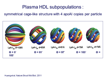
To better understand the interactions of apoA-I in the HDL particle structure, the next step was to take the 5 subpopulations of HDL particles discussed in Figure 4, the HDL2s and HDL3s, and separate out all of the particles that contained both apoA-I and apoA-II, leaving only the particles that contained simply apoA-I. These are the particles shown in the Figure.[8]
In this preparation, the HDL particles were now found to contain, not 3, but 4 copies of apoA-I per particle, and these were organized rather like 4 segments in an orange. Thus the Figure shows a schematic that can be imagined as an orange with 4 segments, here colored red, green, blue, and yellow.
The question, clearly, was to understand how both the large HDL (the HDL2, on the far left in the Figure, referred to here as LpA-I2b or -2a) and the small particles (the HDL3, or the 3 right particles in the Figure) could all, from the largest to the smallest, be comprised of 4 copies of apoA-I?
To investigate this, Sean Davidson and his colleagues had the brilliant idea of, in effect, taking the “top of the orange” and the “bottom of the orange” and twisting them in opposite directions. The result was that the size of the “orange” became progressively smaller, until ultimately it had gone from a particle the size of HDL2b down to size HDL3c, all the while maintaining the basic structure of 4 apoA-I copies per particle.
This was a very important discovery, of course, because it meant that the amount of lipid (as opposed to protein) that was exposed on the surface of the particle is much greater in the large than in the small particles – and this, of course, has major implications for HDL metabolism and function, as we will come back to in the following schematics.
J Clin Lipidol. 2011; 5(6).References
[8]Huang R, Gangani RA, Silva D, et al. Apolipoprotein A-I structural organization in high- density lipoproteins isolated from human plasma. Nature Stuct Mol Biol 2011; 18: 416- 422.