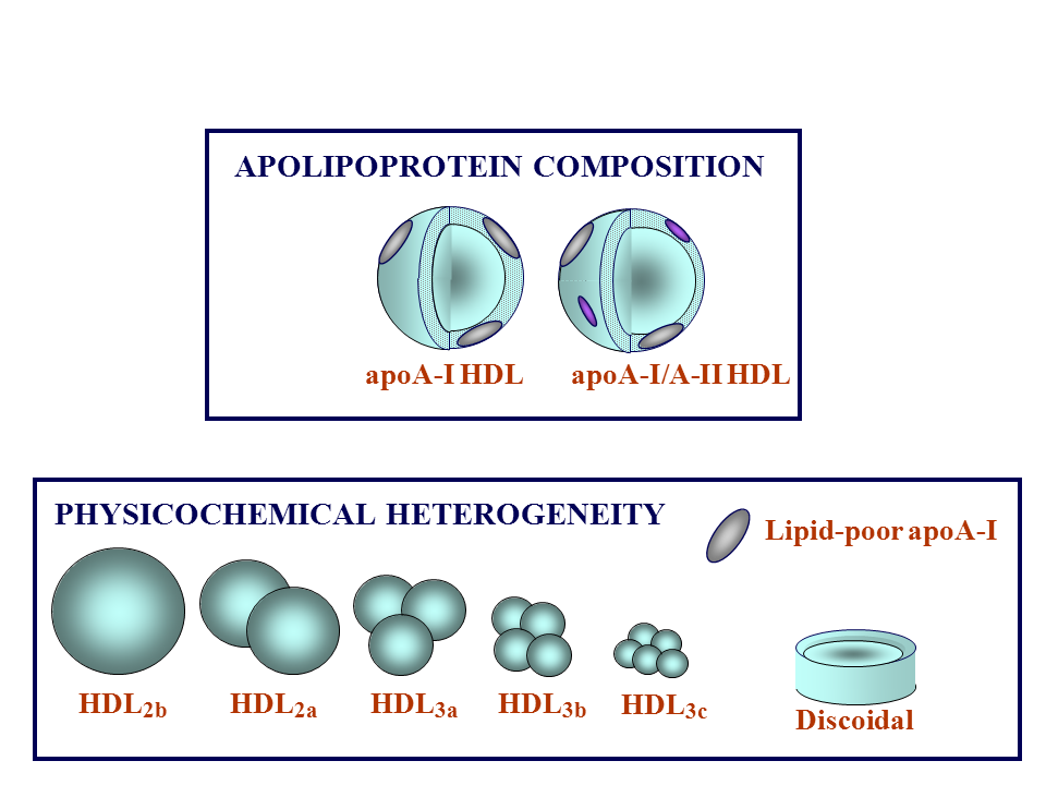
This Figure shows the different types of HDL particles that are in the circulation. Some of those particles are simply lipid-poor forms of apoA-I, and these primarily correspond to forms that are produced by the liver and the intestine and secreted into the circulation. The discoidal forms shown in the Figure represent apoA-I that has gained more lipid, in part by effluxing lipid from other cells (eg, macrophages), and in these discoidal structures, apoAI assumes what is called a “belt” structure – as discussed in the following Figures.
Once ingested into one of these discoidal HDL particles, the free cholesterol becomes esterified by the enzyme lecithin cholesterol acyltransferase (LCAT), to form cholesterol ester. This cholesterol ester is a neutral lipid, and as such it constitutes a core within HDL particles. As LCAT acts on more free cholesterol that is effluxed into the nascent HDL particle, these nascent discoidal HDL particles gradually become larger, spherical particles surrounding a core of LCAT-esterified (hydrophobic) cholesterol.
As these HDL particles continue to grow in size, they form a family, or spectrum, of particle sizes, beginning with what is labeled as HDL3 or HDL3c (the smallest) progressing in size up to HDL2 or HDL2b (the largest). This size nomenclature is simply one of several different ones that are used to identify different HDL subpopulations; in addition to size, which is determined when the particles are isolated from serum by ultracentrifugation, these different particles can equally well be isolated electrophoretic methods.
There are 2 major structural proteins in HDL. One of them, already discussed, is apoAI; the second protein is called apoA-II. Some HDL particles contain only apoA-I, and others, about a third of all the HDL particles, contain both apoA-I and apoA-II.
In all of the preceding discussion we have identified HDL particles with apoA-I – so what, exactly, is the apoA-II identified in the Figure? ApoA-II is a smaller protein, about half or one third the size of ApoA-I, and because the ApoA-II contains a cysteine residue, apoA-I is able to form a bridge of cysteine, and that disulfide bond links to apoA-II monomers to form a dimer. So in the ApoA-I/ApoA-II HDL particles there is a significant role for ApoA-II in establishing the structure of the final HDL particle.
Finally, when talking about the HDL2 and HDL3 subpopulations, it is important to understand that all of those subpopulations consist of a mixture of particles containing either ApoA-I alone or both ApoA-I and ApoA-II – and now we start to see how complex the spectrum of HDL particle subpopulations truly is. This is particularly important because compared with the low-density lipoprotein (LDL) particles, the spectrum of HDL particles is far more complex and even today, HDL biology is not completely understood.
J Clin Lipidol. 2011; 5(6).