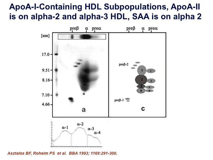
This Figure shows the results when serum lipids are passed through a 2-dimensional (2-D) electrophoresis gel. When serum is run through this gel, it separates the lipids from large to small in the vertical dimension and by charge in the horizontal dimension. Then the gels are blotted with an antibody to apoA-I, the major protein in the HDL particles, to specifically highlight the apoA-I-containing particles, ie, the HDL particles. The righthand panel of this gel then reproduces the blots in cartoon form, labeled as the spectrum of HDL particles, from the very small, pre-beta-1 particles to the spectrum of “mature” alpha-HDL particles, alpha-4, -3, -2, and -1, migrating through the gel from small to large, respectively, in size.
J Clin Lipidol. 2011; 5(6).