|
||
|
American Journal of Medicine
|
|
|
||
|
American Journal of Medicine
|
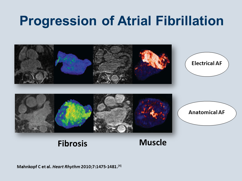
As a disease atrial fibrillation is heterogeneous at many levels. From a cardiac structural perspective (Figure 3) it can be seen to exist at two extremes.[4]
In one case, the atrial myocardium seems normal: the myocardium is preserved; there is little fibrosis; the chambers of the heart are not enlarged; and the ventricles are not dysfunctional. This is described as electrical atrial fibrillation.
At the other extreme, the atrium may be large; the ventricles may be dysfunctional; there may be valvular regurgitation; extensive amounts of fibrosis exist; and atrial myocardium is progressively lost. This extreme is known as anatomical atrial fibrillation.
It is important to perceive these differences, because purely electrical atrial fibrillation probably does not produce thromboembolism, whereas thromboembolism is much more common in patients with anatomical atrial fibrillation. Camm J. Am J Med 2013; published on-line at http://education.amjmed.com/00000.
[4] Mahnkopf C, Badger TJ, Burgon NS, et al. Evaluation of the left atrial substrate in patients with lone atrial fibrillation using delayed-enhanced MRI: implications for disease progression and response to catheter ablation. Heart Rhythm. 2010;7:1475-1481.